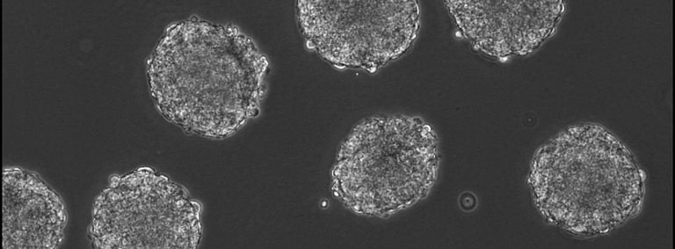
As far as conventional imaging methods go, changes in the tumor microenvironment have consistently evaded detection. However, researchers at the Baylor College of Medicine and Texas Children’s Hospital have developed a new, noninvasive method to measure and monitor the effectiveness of cellular immunotherapies. This new approach to imaging is called “nano-radiomics” and it uses quantitative analysis of nanoparticle contrast–enhanced three-dimensional images to detect tumor response to cellular immunotherapy. Knowing about the responsiveness of the tumor microenvironment to various therapies is becoming more important now that we know that the tumor microenvironment is capable of inhibiting certain immunotherapies. Compared to previous imaging methods, researchers are able to detect texture-based features within tumors as opposed to merely seeing whether a tumor is growing or shrinking. With these new details being revealed to clinicians, we will be better able to enhance cancer treatment and management. To those interested in more detail, here is the article link: https://www.genengnews.com/news/tumor-microenvironment-analyzed-by-new-nano-radiomics-approach/?utm_medium=newsletter&utm_source=GEN+Daily+News+Highlights&utm_content=01&utm_campaign=GEN+Daily+News+Highlights_20200714&oly_enc_id=6133D5788901G9A
Kaitlyn Grayson’s Blog
The journey of a researcher in training
